2006
Prof. Frank Hadley Collins, Dir., Cntr. for Global Health and Infectious Diseases, Univ. of Notre Dame
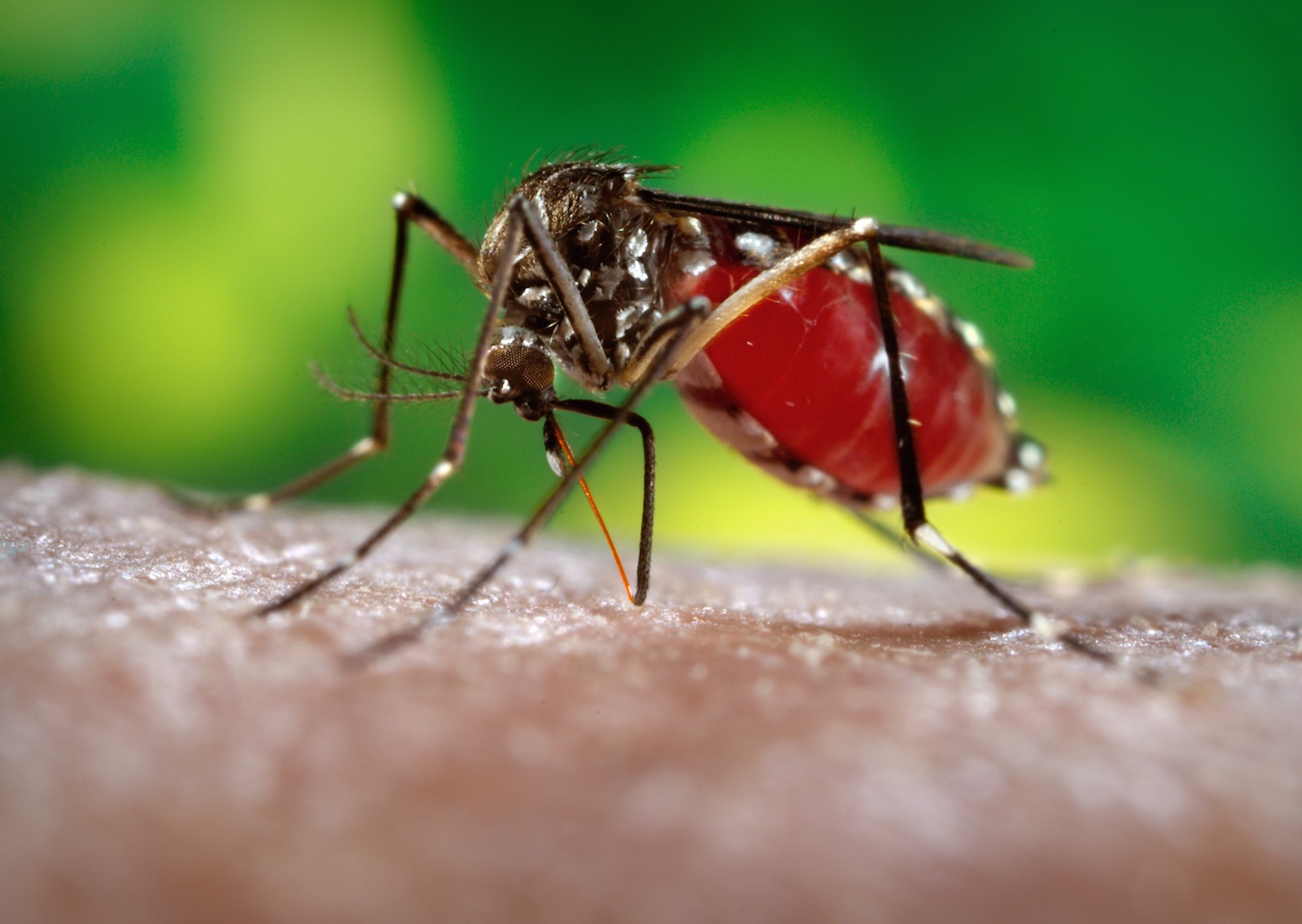
This 2006 photograph depicted a female Aedes aegypti mosquito while she was in the process of acquiring a blood meal from her human host, who in this instance, was actually the biomedical photographer, James Gathany, here at the Centers for Disease Control. You’ll note the feeding apparatus consisting of a sharp, orange-colored “fascicle”, which while not feeding, is covered in a soft, pliant sheath called the “labellum”, which retracts as the sharp stylets contained within pierce the host’s skin surface, as the insect obtains its blood meal. The orange color of the fascicle is due to the red color of the blood as it migrates up the thin, sharp translucent tube.
The first reported epidemics of Dengue (DF) and dengue hemorrhagic fever (DHF) occurred in 1779-1780 in Asia, Africa, and North America. The near simultaneous occurrence of outbreaks on three continents indicates that these viruses and their mosquito vector have had a worldwide distribution in the tropics for more than 200 years. During most of this time, DF was considered a mild, nonfatal disease of visitors to the tropics. Generally, there were long intervals (10-40 years) between major epidemics, mainly because the introduction of a new serotype in a susceptible population occurred only if viruses and their mosquito vector, primarily the Aedes aegypti mosquito, could survive the slow transport between population centers by sailing vessels.